生物/神经研究技术——Tissue Clearance组织透明化
从2012/2013年3DISCO和CLARITY发表以来,各种组织透明化方法层出不穷,各个方案的侧重点不同,但几乎都是在以下几个方面做改进:

组织透明化的兴起,和两个东西关系紧密,一个是光片显微镜的应用(light-sheet microscopy),一个是神经生物学研究的发展。
光片显微镜在2008年第一次提出,广泛应用于3D生物样本的成像,具有速度快、损耗小等优点;但是生物样本有个大问题就是光散射严重,这限制了光片显微镜的应用,组织透明化就可以解决这个问题。去除/溶解组织中的脂质,使得组织变透明、散射减小,对3D成像很有帮助。
神经生物学近年来受到广泛关注。神经生物学研究方法基于两大类,在生物体上的研究,离体模型的研究。前者常常受到现实因素的限制,因此形形色色的离体模型被提出,提取出的组织(切片),类器官,往往相比2D模型更接近于生物体,因此在很多情况下被认为是更好的模型,这些模型的研究需要3D成像技术,因此也促进了组织透明化的研究。
组织透明化,很好理解,顾名思义,就是把组织变透明,简单来说其做法就是把组织中的脂质去掉/溶解。
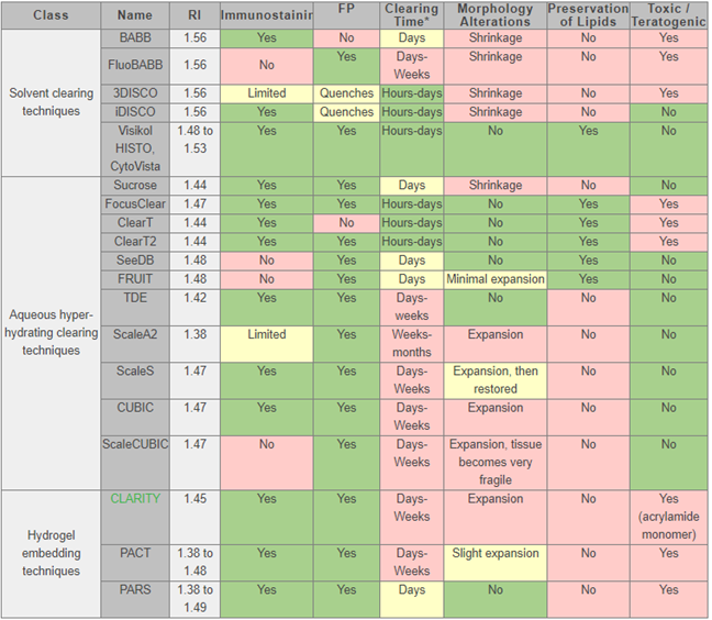
大体上有三类方法:

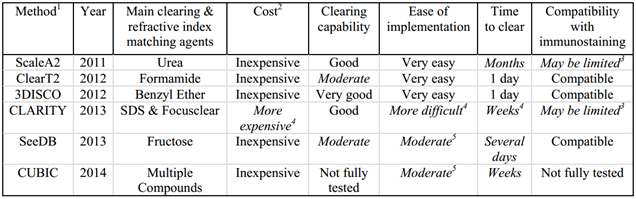
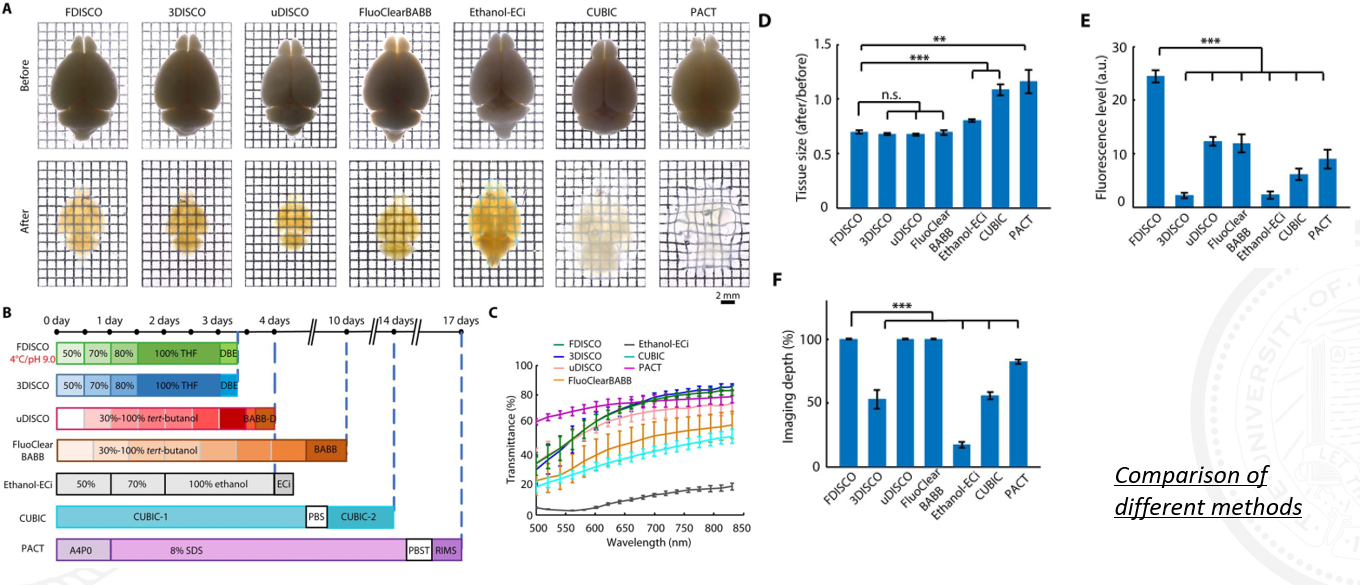
有人对这些方法做了对比:


论文报道的和商用的方法我整理了一些我感兴趣的/引用较高的,附上参考文献和链接:
CLARITY
Chung K, Wallace J, Kim S Y, et al. Structural and molecular interrogation of intact biological systems[J]. Nature, 2013, 497(7449): 332-337.
Chung K, Deisseroth K. CLARITY for mapping the nervous system[J]. Nature methods, 2013, 10(6): 508.
https://logosbio.com/tissue-clearing_3d-imaging/intro
https://www.abcam.com/protocols/clarity-protocol
Visikol HISTO, the first commercially available, easy-to-use, non-destructive and rapid tissue clearing technology
https://www.abcam.com/kits/tissue-clearing-methods-and-reagents
https://visikol.com/products/visikol-histo/
CUBIC
Susaki E A, Tainaka K, Perrin D, et al. Whole-brain imaging with single-cell resolution using chemical cocktails and computational analysis[J]. Cell, 2014, 157(3): 726-739.
Susaki E A, Tainaka K, Perrin D, et al. Advanced CUBIC protocols for whole-brain and whole-body clearing and imaging[J]. Nature protocols, 2015, 10(11): 1709.
SWITCH
Murray E, Cho J H, Goodwin D, et al. Simple, scalable proteomic imaging for high-dimensional profiling of intact systems[J]. Cell, 2015, 163(6): 1500-1514.
http://www.chunglabresources.com/sw1/
3/i/u/v/FDISCO
Ertürk A, Becker K, Jährling N, et al. Three-dimensional imaging of solvent-cleared organs using 3DISCO[J]. Nature protocols, 2012, 7(11): 1983.
Renier N, Wu Z, Simon D J, et al. iDISCO: a simple, rapid method to immunolabel large tissue samples for volume imaging[J]. Cell, 2014, 159(4): 896-910.
Pan C, Cai R, Quacquarelli F P, et al. Shrinkage-mediated imaging of entire organs and organisms using uDISCO[J]. Nature methods, 2016, 13(10): 859.
Cai R, Pan C, Ghasemigharagoz A, et al. Panoptic vDISCO imaging reveals neuronal connectivity, remote trauma effects and meningeal vessels in intact transparent mice[J]. BioRxiv, 2018: 374785.
Qi Y, Yu T, Xu J, et al. FDISCO: Advanced solvent-based clearing method for imaging whole organs[J]. Science advances, 2019, 5(1): eaau8355.
SeeDB/SeeDB2
Ke M T, Fujimoto S, Imai T. SeeDB: a simple and morphology-preserving optical clearing agent for neuronal circuit reconstruction[J]. Nature neuroscience, 2013, 16(8): 1154.
Ke M T, Nakai Y, Fujimoto S, et al. Super-resolution mapping of neuronal circuitry with an index-optimized clearing agent[J]. Cell reports, 2016, 14(11): 2718-2732.
FOCM
Zhu X, Huang L, Zheng Y, et al. Ultrafast optical clearing method for three-dimensional imaging with cellular resolution[J]. Proceedings of the National Academy of Sciences, 2019, 116(23): 11480-11489.
COMPARISON
https://visikol.com/tissue-clearing-comparison/
很有意思的是,有一些基于溶剂的方法,尽管开发者并不都是属于同一课题组,却都沿用了一样格式的名字:XDISCO
做了个对比:
每个方法都是基于溶解脂质,但是在荧光保存,清除时间,免疫荧光/荧光蛋白侧重上都不同。

参考资料:
附在文中

 浙公网安备 33010602011771号